
Plasma Cell Myeloma : Radiologic Evaluation


Comments:
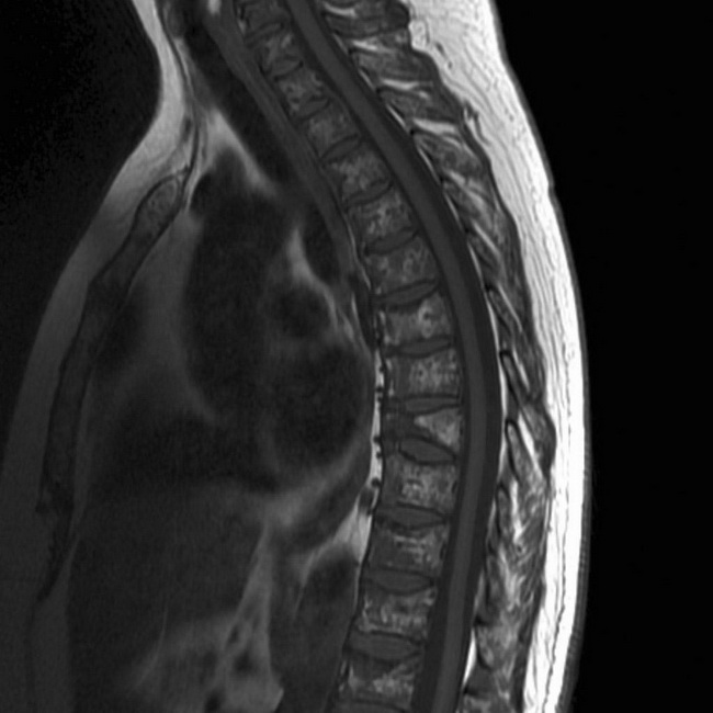
Given that the most common presenting symptom is bone pain induced by lytic lesions, radiology plays a key role in both the diagnosis as well as the management of plasma cell myeloma. A skeletal survey (i.e. a series of radiographs systematically performed to cover the entire skeleton) is essential in making the diagnosis, assessing the response to treatment, as well as pre-empting mechanical complications such as pathologic fractures. Computed tomography scans and magnetic resonance imaging (MRI) are more sensitive than plain radiographs in detecting small bone lesions as well as extramedullary plasmacytomas. This MRI of the thoracic spine demonstrates extensive patchy regions of bone marrow replacement by the tumor (normal fatty marrow is bright on both T1 and T2 - abnormal marrow is dark) associated with multiple superior endplate crush fractures. Features are consistent with plasma cell myeloma (pathologically confirmed). Case courtesy of A.Prof Frank Gaillard, Radiopaedia.org. From the case rID: 9968