
Paget Disease of Bone : Radiology


Comments:
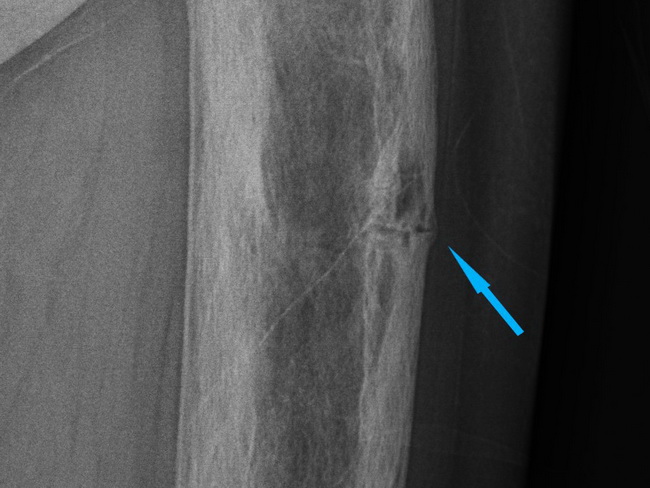
Paget Disease of Bone (PDB) - Imaging Studies: Enlarged annotated view of plain radiograph shown in the previous image. The arrow shows transverse insufficiency fracture (aka banana fracture) at the lateral aspect of the cortex of the femur. The patient presented four years later with a complete transverse fracture at the same site (see next image). About 10-30% of PDB patients sustain pathologic fractures which may be incomplete initially, traversing the outer border of the cortex of bowed bones. These fissure fractures usually occur in the weight bearing bones and are at increased risk of complete transverse fracture (as happened in this case; see next image). Case courtesy of Dr Salman S. Albakheet, Radiopaedia.org. From the case rID: 80359