
Paget Disease of Bone : Microscopic

slide 26 of 48


Comments:
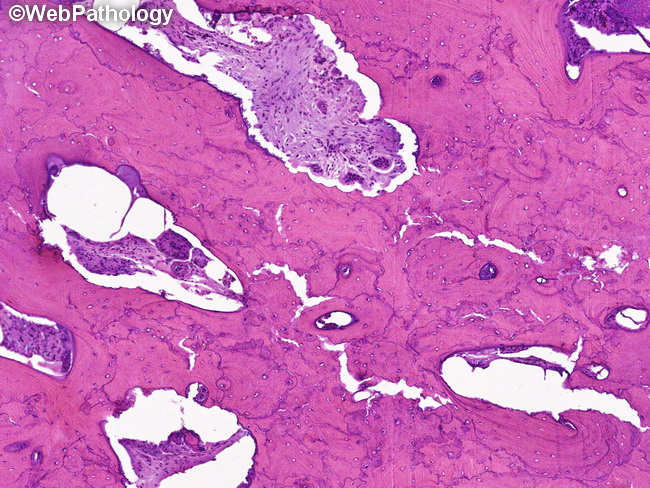
Paget Disease of Bone (PDB) - Mixed Osteolytic & Osteoblastic Phase: This image shows thick, irregular, plate-like bony trabeculae with several resorption cavities. The marrow space is filled with fibrovascular tissue containing scattered osteoclasts. Osteoblasts are not seen here, although they were prominent in other sections from this case. Note the prominent, irregular blue cement lines, creating a mosaic pattern as well as microcracks in the trabeculae. These changes are non-specific and pagetoid bone may be seen with any condition with high bone turnover such as metastatic carcinoma and osteosarcoma. Correlation with imaging studies is critical.
slide 26 of 48