
Myositis Ossificans - Morphology

slide 12 of 24


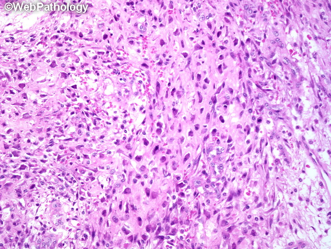
Comments:
Microscopic Features: At the interface between the innermost and intermediate zones of myositis ossificans there is an admixture of fibroblasts and osteoblasts and appearance of poorly-defined trabeculae with variable amounts of osteoid. Hypercellular chondroid foci may be present in some cases. Taken out of context, such foci may be mistaken for chondrosarcoma or chondroblastic osteosarcoma.
slide 12 of 24