
Chordoma : Cytology

slide 18 of 51


Comments:
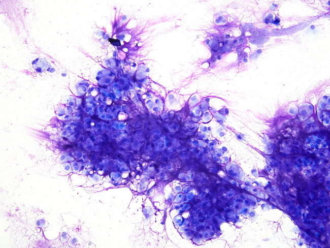
Chordoma - Cytology: Smears typically show tumor cells, singly and in clusters, surrounded by copious amounts of metachromatic watery myxoid matrix with a fibrillar appearance as shown here. The cells are large and have multivacuolated cytoplasm (physaliphorous cells). Signet ring cell forms may be present. The nuclei are large but bland. Image courtesy of: Syed Z. Ali, MD; used with permission.
slide 18 of 51