
Osteoid Osteoma : Radiographic Features

slide 7 of 13


Comments:
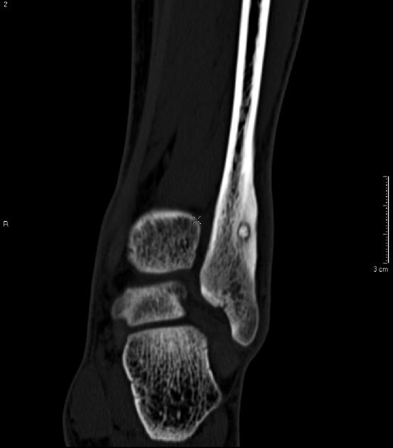
A computed tomographic scan of the knee showing osteoid osteoma in the lower part of fibula. This young male presented with a history lateral ankle pain. Case produced with permission, courtesy of Dr. Frank Gaillard. Radiopaedia. Complete case is here.
slide 7 of 13