
Chondromyxoid Fibroma

slide 8 of 17


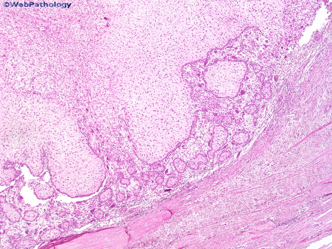
Comments:
As the name suggests, chondromyxoid fibroma shows chondroid, myxomatous, and fibrous zones. The tumor has a lobular growth pattern with lobules of varying sizes as seen in this image. The lobules have a hypocellular center with condensation of tumor cells at the periphery.
slide 8 of 17