
Chondromyxoid Fibroma

slide 15 of 17


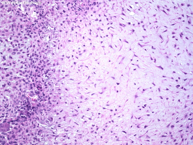
Comments:
Hypocellular myxoid areas near the center of the lobule give way to more cellular (chondroblastoma-like areas) at the periphery. The nuclei of the tumor cells are round, oval, spindle-shaped or stellate-shaped. The most important differential diagnosis is myxoid chondrosarcoma. Features supporting chondrosarcoma include liquefactive changes in matrix, permeation of surrounding bone, hypercellularity, and malignant features on radiographs.
slide 15 of 17