
Chondromyxoid Fibroma

slide 11 of 17


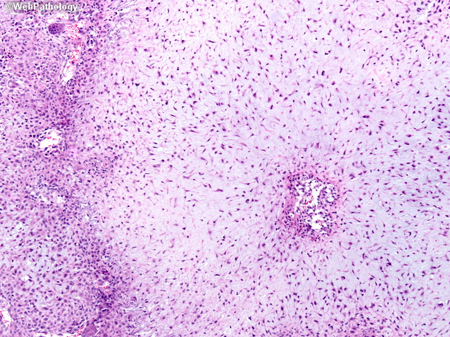
Comments:
The hypocellular center of a lobule is seen on the rightt. The hypercellular periphery on the left shows scattered giant cells and numerous mononuclear cells which mimic the appearance of a chondroblastoma.
slide 11 of 17