
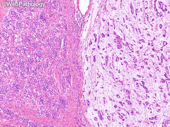
Yolk Sac Tumor : Glandular


Comments:
Yolk sac tumor shows a variety of architectural patterns that are admixed in varying proportions. Glandular/alveolar pattern (one-third of cases): Columnar tumor cells form simple tubules with round/oval lumen or complex anastomosing glandular structures. The glands may merge with the vesicles of polyvesicular vitelline pattern or may be present in a myxomatous, microcystic, or solid background. Some cases show subnuclear vacuoles similar to those seen in secretory endometrium. The glands often show enteric differentiation but lack a smooth muscle layer. Pure glandular yolk sac tumor is more common after chemotherapy and is therefore seen more frequently in metastases and late recurrences. The image shows pure glandular pattern in a prepubertal yolk sac tumor. Note the immature seminiferous tubules composed mostly of Sertoli cells in the left half of the image.