
Invasive Ductal CA : Mitotic Rate


Comments:
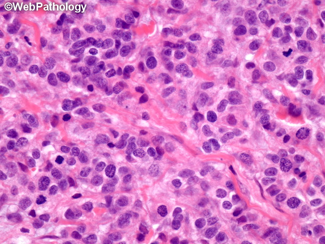
Mitotic rate is one of the most important prognostic features in the modified Scarff-Bloom-Richardson histologic grading system. Mitotic figures are counted with 40x objective in 10 consecutive fields (field area of 0.196 sq.mm) with invasive tumor. The selected area should be the most cellular invasive region at the periphery of the tumor where mitotic activity tends to be the highest. It should be away from necrosis, inflammation, areas of calcification and large blood vessels. It is important to employ standardized counting methods and use fixed field size when assessing mitotic activity to ensure reliability and reproducibility of results. There are tables available listing mitotic rates per 10 HPF for different scores and for different microscopes. Conversion factors have been calculated to convert mitotic counts per 10 HPF to counts per sq.mm. In the modified Scarff-Bloom-Richardson system, scoring for mitotic figures is done as follows: 0-7 mitoses/10HPF (score 1); 8-14 mitoses/10HPF (score 2); and 15 or more mitoses/10HPF (score 3). Mitotic activity is generally underestimated in needle core biopsy specimens from invasive carcinomas. The use of mitosis-specific antibody such as PHH3 and MIB1 in needle core biopsy specimens may give a better correlation with mitotic count in the excised specimens. The image shows invasive ductal carcinoma with intermediate-grade nuclei (score 2), no tubule formation (score 3) and 10 mitoses/10HPF (score 2). This field shows 3 mitotic figures.