
Prostate Cancer

slide 3 of 55


Comments:
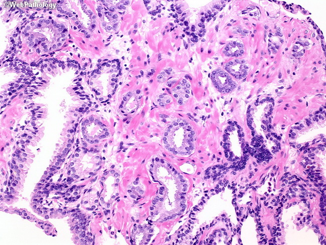
The image shows about a dozen small glands with round or oval lumens infiltrating in between obviously benign larger glands. The smaller atypical glands are lined by a single layer of cells and show nuclear enlargement with prominent nucleoli. Basal cells are not visible. In contrast, the larger glands have complex branching lumens and many of them contain clearly visible basal cells with darker nuclei. See next image for high molecular weight cytokeratin immunostain.
slide 3 of 55