
Heterotopic Pancreas : Differential


Comments:
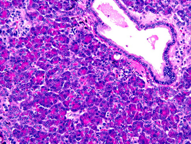
Differential Diagnosis: Grossly, heterotopic pancreas can resemble gastrointestinal stromal tumors, lipomas, and leiomyomas. Microscopically, it should be distinguished from the following entities: Infiltrating well-differentiated pancreatic adenocarcinoma: This is the most important differential diagnostic consideration. Features favoring heterotopic pancreas include: lobular architecture resembling normal pancreas, absence of cytologic atypia, lack of desmoplastic stroma, and presence of smooth muscle bundles within various components. Adenomyoma: Heinrich type III pancreatic heterotopia consists of only ductal elements surrounded by smooth muscle fascicles and can mimic adenomyoma. Pancreatic Acinar Metaplasia: Gastric biopsies from cardia frequently show pancreatic acinar metaplasia. However, these are microscopic foci, do not form a mass, and are usually associated with inflammatory conditions such as reflux esophagitis. Paneth Cell Metaplasia: Chronic atrophic gastritis and autoimmune gastritis may be associated with intestinal metaplasia and Paneth cell metaplasia. Paneth cells have large bright orange-red refractile granules. Pancreatic acinar cells have fine basophilic granules at the base and pink granules at the apex. Neuroendocrine Tumor (NET): Heinrich type IV pancreatic heterotopia is composed of only islet cells and may mimic neuroendocrine tumor. However, it is composed of small nests of islet cells in the submucosa and muscularis propria. Unlike NET, there is no mass formation and there is no stromal response. About this image: Heterotopic pancreatic tissue in stomach, Heinrich type I (all cell types present; the most common subtype). The image shows pancreatic acini, a duct, and islet cells (upper left). Image courtesy of: Sanjay Mukhopadhyay, MD.