
Gallbladder : Adenomyoma

slide 9 of 14


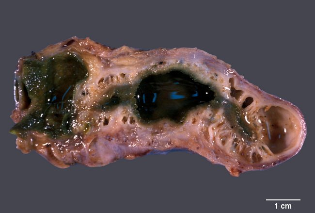
Comments:
Adenomyoma consists of cystically dilated glands forming a small mass or nodule in the wall of the gallbladder. It has also been referred to as adenomyomatous hyperplasia or nodule. When the process is diffuse (uncommon; shown in this image), it is called adenomyomatosis. Macroscopically, the nodule shows cystic spaces creating a sieve-like appearance. The most common location is fundus. Image courtesy of: Dr. Ibrahim Zardawi; used with permission.
slide 9 of 14