
Infantile Hemangioendothelioma : Liver

slide 6 of 10


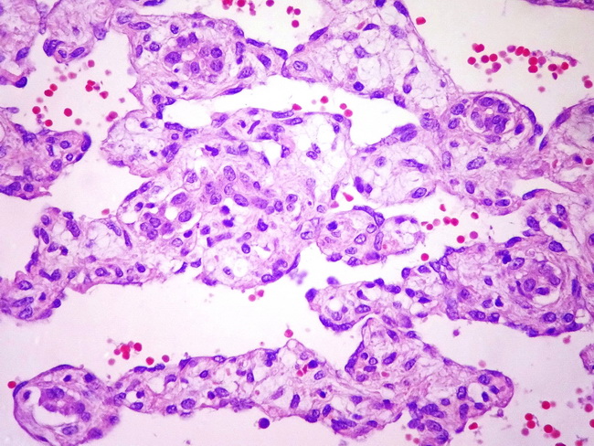
Comments:
Infantile hemangioendothelioma of liver: The vascular lumina contain red blood cells and are lined by bland endothelial cells. Entrapped small bile ducts can be seen in the supporting stroma. Image courtesy of: Mohammad Adib Houreih, MD; Dept. of Pathology, Tishreen University, Lattakia, Syria.
slide 6 of 10