
Fibrolamellar Carcinoma of Liver

slide 59 of 65


Comments:
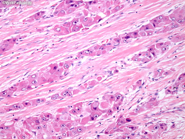
High magnification of FLC reveals cords of large, polygonal tumor cells with large nuclei, prominent nucleoli, and abundant granular eosinophilic cytoplasm. Dense bands of lamellar fibrosis separate the tumor cell cords. This variant should be distinguished from the scirrhous pattern of conventional HCC, which lacks diffuse parallel arrays of dense fibrosis and the tumor cells have less granular, eosinophilic cytoplasm.
slide 59 of 65