
Focal Nodular Hyperplasia : Additional Image
Home
 Gastrointestinal
Liver
Liver Tumors & Tumor-like Lesions - I
Focal Nodular Hyperplasia : Additional Image
Gastrointestinal
Liver
Liver Tumors & Tumor-like Lesions - I
Focal Nodular Hyperplasia : Additional Image

slide 48 of 63


Comments:
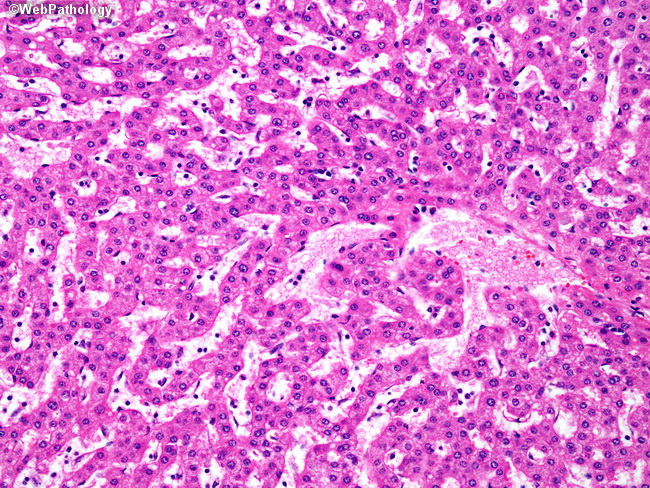
The liver cell plates in focal nodular hyperplasia (FNH) are orderly and composed of mature, uniform hepatocytes with occasional mild cytologic atypia, enlarged nuclei, and mild hyperchromasia. Mitotic activity is very rare. FNH shows variable sinusoidal dilatation and scattered Kupffer cells (CD68+). With FNHs, there is no risk of malignant transformation and very low risk of complications such as hemoperitoneum. Many cases are left alone if they can be confidently diagnosed with imaging studies.
slide 48 of 63