
Focal Nodular Hyperplasia : Additional Image
Home
 Gastrointestinal
Liver
Liver Tumors & Tumor-like Lesions - I
Focal Nodular Hyperplasia : Additional Image
Gastrointestinal
Liver
Liver Tumors & Tumor-like Lesions - I
Focal Nodular Hyperplasia : Additional Image

slide 46 of 63


Comments:
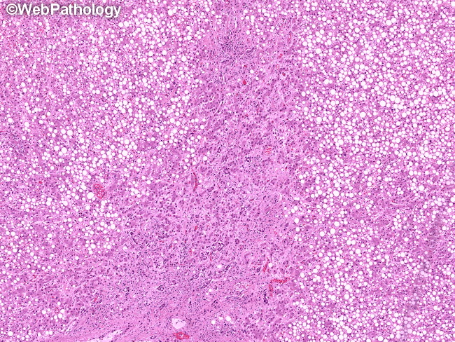
The hepatocytes in focal nodular hyperplasia are relatively uniform with variable amounts of cytoplasmic glycogen, lipid (as seen here), bile, lipofuscin, iron pigment, or Mallory bodies. Occasionally, there is focal cytologic atypia, with enlarged nuclei, mild hyperchromasia, and conspicuous nucleoli. Mitotic activity is extremely rare and, if present, suggests the possibility of hepatocellular carcinoma. The center of this image shows a thick fibrous band with prominent ductular reaction.
slide 46 of 63