
Focal Nodular Hyperplasia : Microscopic
Home
 Gastrointestinal
Liver
Liver Tumors & Tumor-like Lesions - I
Focal Nodular Hyperplasia : Microscopic
Gastrointestinal
Liver
Liver Tumors & Tumor-like Lesions - I
Focal Nodular Hyperplasia : Microscopic

slide 38 of 63


Comments:
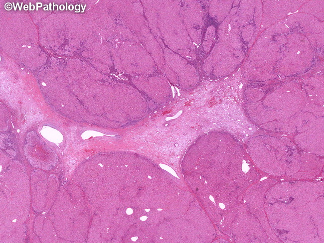
Microscopic Features: At low magnification, focal nodular hyperplasia shows multiple nodules separated by thick fibrous bands originating from the central scar. The central scar, which may be absent in some cases, contains one or more dystrophic thick-walled blood vessels (without accompanying bile ducts) and numerous arterioles. The vessels may show intimal and smooth muscle hyperplasia, subintimal fibrosis, and thrombosis. The darker areas between the nodules represent chronic inflammatory infiltrate in the fibrous bands.
slide 38 of 63