
Focal Nodular Hyperplasia : Imaging
Home
 Gastrointestinal
Liver
Liver Tumors & Tumor-like Lesions - I
Focal Nodular Hyperplasia : Imaging
Gastrointestinal
Liver
Liver Tumors & Tumor-like Lesions - I
Focal Nodular Hyperplasia : Imaging

slide 37 of 63


Comments:
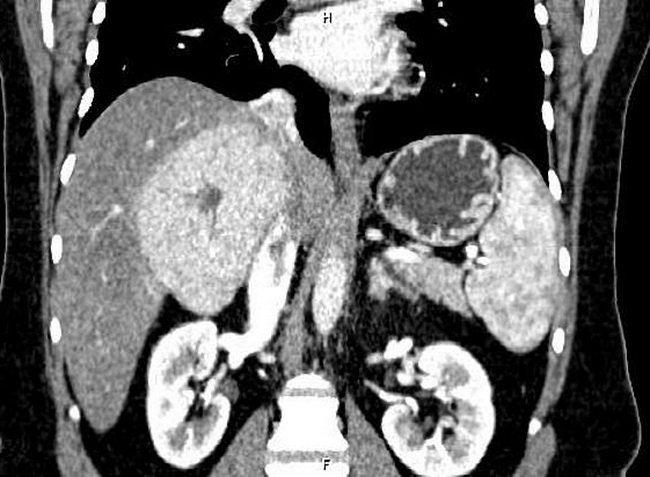
The diagnosis of focal nodular hyperplasia (FNH) of liver can be accurately done in most cases with contrast-enhanced CT or MRI study. This CT with contrast (coronal view) shows a large, partially exophytic mass in the liver with bright contrast enhancement in the arterial phase. The darker non-enhancing area within the lesion is the central scar. Case courtesy of Dr Mohammad Taghi Niknejad, Radiopaedia.org. From the case rID: 64211
slide 37 of 63