
Focal Nodular Hyperplasia : Gross Pathology
Home
 Gastrointestinal
Liver
Liver Tumors & Tumor-like Lesions - I
Focal Nodular Hyperplasia : Gross Pathology
Gastrointestinal
Liver
Liver Tumors & Tumor-like Lesions - I
Focal Nodular Hyperplasia : Gross Pathology

slide 32 of 63


Comments:
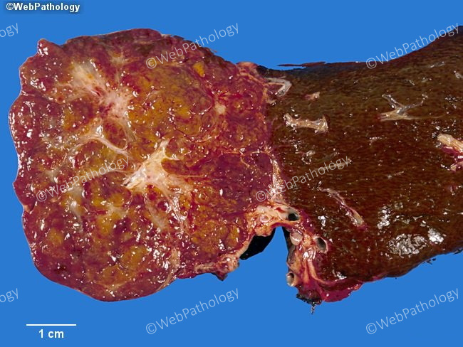
Gross Pathology: Focal nodular hyperplasia (FNH) is usually <5 cm but there is great size variation. Most FNH >1 cm have central scar which consists of fibrovascular tissue (not dense collagen). The specimen shown here is partially exophytic and has multiple stellate scars. A rare telangiectatic variant of FNH was previously described that lacks a central fibrous zone and may resemble vascular lesions (such as hemangioma) or peliosis hepatis. This lesion is now reclassified as an inflammatory hepatocellular adenoma.
slide 32 of 63