
Focal Nodular Hyperplasia : Gross Pathology


Comments:
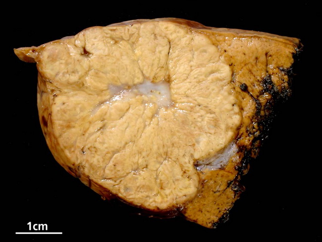
Gross Pathology: Focal nodular hyperplasia (FNH) is subcapsular, solitary, well-circumscribed (but unencapsulated), firm, coarsely nodular lesion with a central stellate scar. It is multifocal in about 20% of cases. Due to its multinodular appearance, it resembles macronodular cirrhosis. The size ranges from a <1 cm to >10 cm. It is yellow-tan or gray and lighter in color than adjacent liver parenchyma. Fibrous septa radiate outwards from the depressed central scar and divide the lesion into incomplete nodules. Central scar is seen in 60-70% of cases but it may be poorly developed or absent in some cases. FNH may be partially exophytic or even pedunculated. Hemorrhage and necrosis are seen less frequently than in hepatocellular adenoma. The background liver is usually appears normal. Image copyright: pathorama.ch