
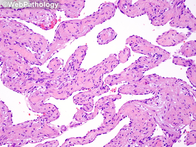
Liver Hemangioma : Microscopic Features
Home
 Gastrointestinal
Liver
Liver Tumors & Tumor-like Lesions - I
Liver Hemangioma : Microscopic Features
Gastrointestinal
Liver
Liver Tumors & Tumor-like Lesions - I
Liver Hemangioma : Microscopic Features

slide 18 of 63


Comments:
Microscopic Features: Cavernous hemangiomas of the liver are composed of dilated, anastomosing, thin-walled vascular channels lined by flattened endothelium with no cytologic atypia or mitotic activity. The channels are supported by thin fibrous septa which often contain mast cells.
slide 18 of 63