
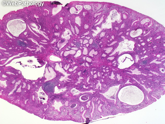
Juvenile Polyp : Microscopic

slide 11 of 45


Comments:
Microscopically, the surface of a juvenile polyp shows ulceration and granulation tissue along with regenerating epithelium which may be mistaken for dysplasia. The cystic spaces are formed by dilated glands containing abundant mucin and inflammatory debris. The glands are separated by an expanded edematous lamina propria containing inflammatory cells.
slide 11 of 45