
Tubulovillous Adenoma

slide 44 of 113


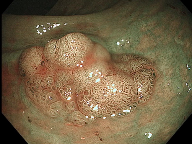
Comments:
Endoscopic view of tubulovillous adenoma of rectum using Narrow-band Imaging (NBI). As previously explained (slide 6), the light source is switched to blue light (415 nm) or green light (540 nm). Due to peak absorption by hemoglobin at these wavelengths, the blood vessels appear darker and vascularity of the lesion is clearly delineated. Endoscopists use NBI in distinguishing between non-neoplastic and neoplastic lesions.
slide 44 of 113