
Mucinous Cystadenoma

slide 5 of 33


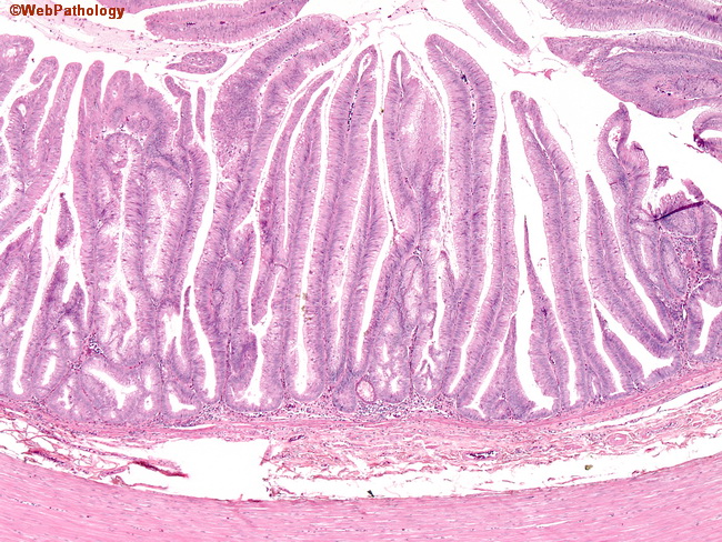
Comments:
Low power view shows long villous processes lined by orderly mucinous epithelium. The low-grade cytology is evident even at this magnification. Appendix involved by a mucinous cystadenoma may show several secondary changes such as hyalinization and fibrosis of the wall, thinning of the wall, disappearance of the lymphoid follicles, ulceration, and dystrophic calcification.
slide 5 of 33