
Barrett Esophagus : Microscopic


Comments:
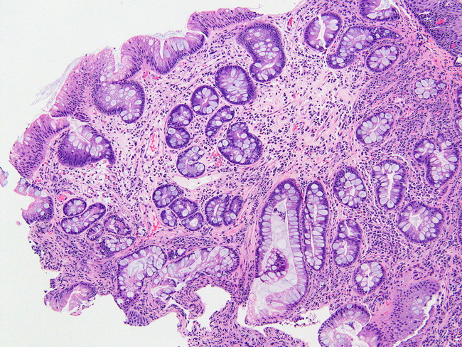
Microscopic Features: For the diagnosis of Barrett esophagus (BE), the guidelines provided by the American College of Gastroenterology require endoscopically abnormal areas in the distal esophagus to show unequivocal intestinal-type epithelium with goblet cells (intestinal metaplasia). In some countries, such as the United Kingdom and Japan, the presence of gastric cardiac-type epithelium which consists solely of mucus-secreting cells is considered sufficient for the diagnosis of BE. The columnar epithelium shows tall/elongated cells with basally oriented nuclei, while the goblet cells are round with pale blue cytoplasm (acid mucin) that pushes the nucleus to the periphery of the cell. Goblet cells may occur singly or in clusters throughout the columnar epithelium. It is important to distinguish goblet cells from pseudogoblet cells to not misdiagnose BE. In contrast to true goblet cells, pseudogoblet cells contain clear to pale pink cytoplasm (neutral mucin) and they are arranged in linear clusters along the epithelium. Additional findings in BE may include scattered inflammatory cells (lymphocytes, plasma cells, neutrophils), pancreatic acinar metaplasia, and the presence of oxyntic or mucous glands. Image courtesy of: Phoenix Bell, MD.