
Gallbladder : Polyarteritis Nodosa


Comments:
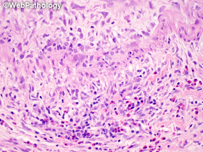
Microscopic Features of Polyarteritis Nodosa in Gallbladder: The classic feature consists of necrotizing vasculitis involving medium sized muscular arteries. There is transmural inflammation of the vessel wall with or without fibrinoid necrosis. The surrounding tissue is devoid of inflammation. The vascular changes are discontinuous with uninvolved segments (skip areas) in between lesions. Fibrinoid necrosis destroys the muscle layers and the internal elastic lamina (best visualized with elastic stains). The inflammatory infiltrate consists of neutrophils, eosinophils, and histiocytes. This is replaced by predominantly lymphocytic infiltrate during the healing phase. Multiple vessels within a specimen often show lesions at varying stages of evolution. Healed lesions show fibrointimal proliferation with narrowing of the vascular lumen. True vasculitis should be distinguished from secondary inflammation of the vessels (bystander phenomenon) that may be seen with generalized acute or chronic cholecystitis. References:1. Odze & Goldblum Surgical Pathology of the GI Tract, Liver, Biliary Tract & Pancreas (2023). 4th edition. Elsevier Saunders.2. Burt A, Ferrell L, Hubscher S. MacSween's Pathology of the Liver (2024). 8th Edition, Elsevier.3. Goldblum, J. R. et al (2018). Rosai and Ackerman's Surgical Pathology. 11th Edition. Philadelphia, PA. Elsevier.