
Choledochal Cyst : Imaging


Comments:
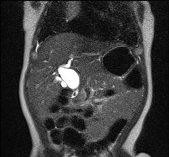
Diagnosis: Diagnosis of choledochal cyst can be made on imaging of the biliary tree with ultrasound, CT, direct contrast studies (ERCP, PTC), nuclear medicine examinations or MRI. Dynamic magnetic resonance cholangiopancreatography is the best imaging study for the evaluation of the biliary tree. This MRCP (coronal T2) shows diffuse fusiform dilation of the common hepatic duct and the common bile duct. Liver and gallbladder appear normal. The findings are consistent with type I choledochal cyst. Case History: The patient was a 5 month old male with history of prematurity and bronchopulmonary dysplasia, and born at 34 weeks gestation. He presented with abdominal pain. There was no history of fever or jaundice. Liver function tests were normal. Case courtesy of Daniel Hyeong Seok Kim, Radiopaedia.org. From the case rID: 154113