
Choledochal Cyst : References


Comments:
References:1. Odze & Goldblum Surgical Pathology of the GI Tract, Liver, Biliary Tract & Pancreas (2023). 4th edition. Elsevier Saunders.2. Burt A, Ferrell L, Hubscher S. MacSween's Pathology of the Liver (2024). 8th Edition, Elsevier.3. Goldblum, J. R. et al (2018). Rosai and Ackerman's Surgical Pathology. 11th Edition. Philadelphia, PA. Elsevier.
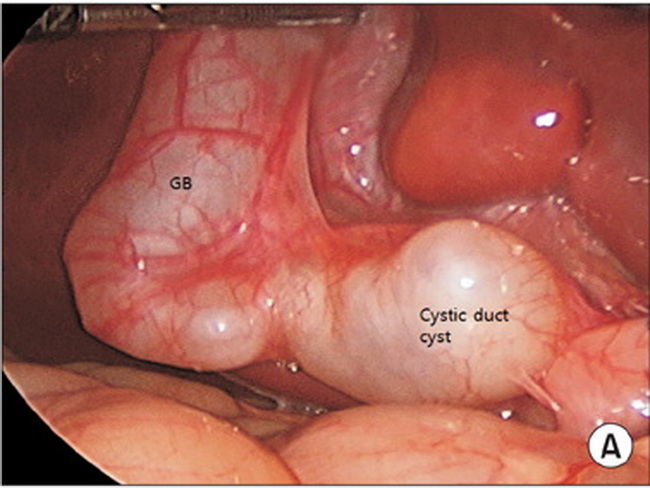
This intraoperative photograph shows dialated cystic duct in a 4 month old boy who presented with abdominal pain and vomiting. Isolated cystic duct cysts are extremely rare and not included in the Todani classification. Some authors refer to them as "type VI," whereas others consider them a subtype of Todani type II. Image source: Joong Kee Youn, Hyejin Kim, Hyun-Young Kim, Sung-Eun Jung. Isolated cystic duct cyst with associated stones in a 4-month-old boy. Annals of Surgical Treatment and Research 2016; 90(6): 350-352. Image used under: Creative Commons Attribution NonCommercial License.