
Gallbladder Carcinoma in Hyalinizing Cholecystitis


Comments:
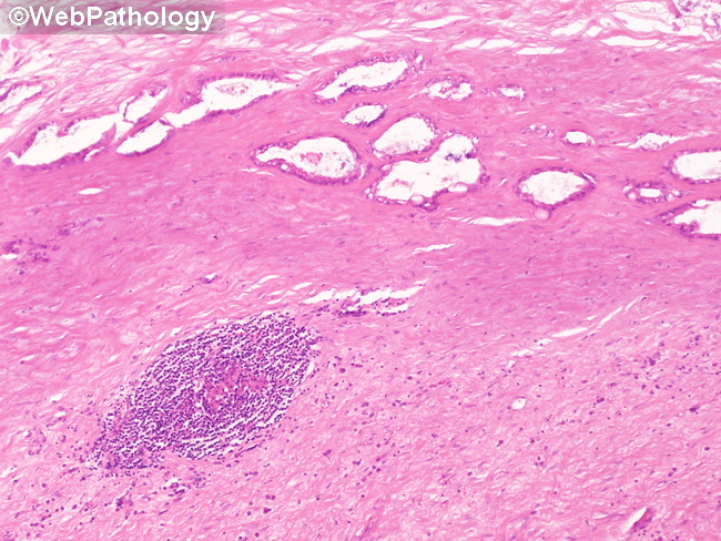
Hyalinizing Cholecystitis-Associated Gallbladder Carcinoma: Carcinomas arising in hyalinizing chronic cholecystitis have subtle features and can be difficult to diagnose correctly. The glands form elongated tubules and have clear cytoplasm, distinct cytoplasmic borders, and small resinoid nuclei. Some glands appear dilated and atrophic and are lined by partially attenuated epithelium. The behavior of hyalinizing cholecystitis-associated carcinomas is more aggressive than ordinary carcinomas as they usually present at advanced stages. Note the features of hyalinizing cholecystitis in this image - dense hyaline fibrosis, low cellularity, and sparse inflammation. In a gallbladder where the wall has been completely effaced by dense hyaline tissue, any glands within the area of fibrosis should raise the concern for carcinoma.