
Xanthogranulomatous Cholecystitis

slide 60 of 74


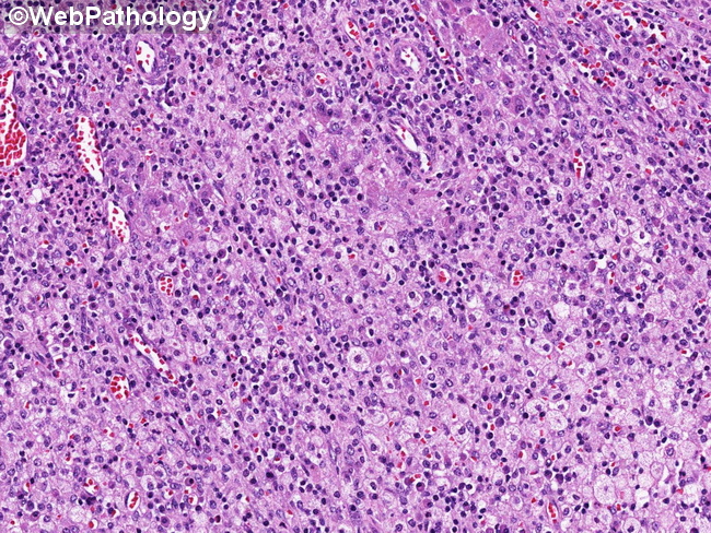
Comments:
Xanthogranulomatous Cholecystitis: The image shows inflammatory infiltrate consisting of lipid-laden macrophages, plasma cells, lymphocytes, scattered neutrophils and eosinophils, in a background with delicate vasculature. The xanthogranulomatous reaction may be focal, with the remainder of the gallbladder showing picture of ordinary chronic cholecystitis with lymphoid follicles.
slide 60 of 74