
Mesenchymal Chondrosarcoma : Clinical Features


Comments:
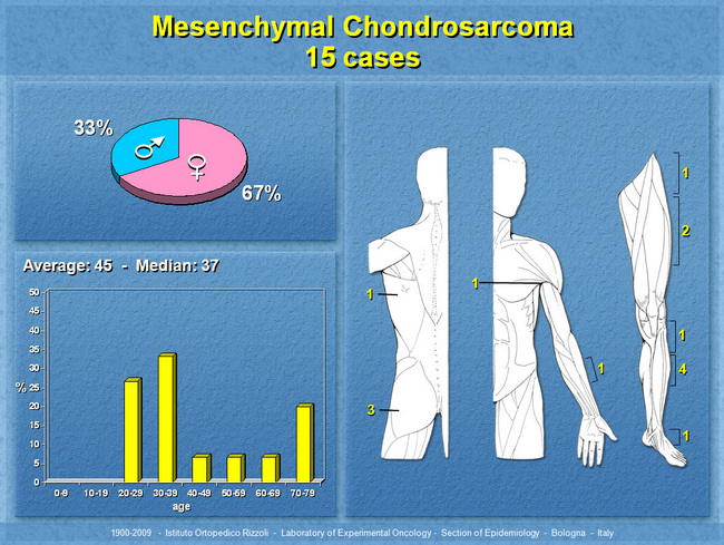
Sites: Mesenchymal chondrosarcoma (MC) accounts for about 2% of chondrosarcomas. It arises in skeletal sites (two-thirds of cases) such as craniofacial bones (common), ribs, vertebrae, pelvic bones, and sometimes diaphysis of long bones (femur, tibia, humerus). One-third of cases involve extraosseous sites like orbit, paraspinal region, meninges, and somatic soft tissues of extremities. Rare cases have involved mediastinum, lungs, or kidneys. Age: It occurs predominantly in adolescents and young adults. Two-thirds of the patients are in 15-35 year age group with a slight female predominance according to some studies. Rare cases occur in young children and neonates. Clinical Features: The usual presentation is with a painful enlarging mass. Some cases are discovered incidentally on imaging studies. Orbital tumors present with exophthalmos, pain, visual disturbances, and headaches. Intracranial and intraspinal tumors cause vomiting, headache, and sensory or motor deficits. Slide courtesy of Piero Picci, M.D., Director, Laboratory of Experimental Oncology, Instituto Ortopedico Rizzoli, Bologna, Italy. Used with permission.