
Syphilitic Lymphadenitis : Steiner Stain
Home
 Hematopathology
Lymph Node (Non-Hematopoietic)
Bacterial Lymphadenitides
Syphilitic Lymphadenitis : Steiner Stain
Hematopathology
Lymph Node (Non-Hematopoietic)
Bacterial Lymphadenitides
Syphilitic Lymphadenitis : Steiner Stain

slide 17 of 17

Comments:
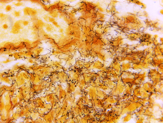
Syphilitic Lymphadenitis: Treponema pallidum can be demonstrated in the lymph node, usually in the walls of capsular vessels with Warthin-Starry or Levaditi stains, immunofluorescent techniques on touch imprints, or by immunohistochemical stains. It can also be detected by PCR on lymph node biopsies or FNA material. Given the variability of histologic findings, the diagnosis is best confirmed with serologic tests for syphilis. This image shows Steiner silver stain on a lymph node section demonstrating numerous, darkly-stained, corkscrew-shaped Treponema pallidum in a case of syphilitic lymphadenitis Image credit: Skip Van Orden/Centers for Disease Control & Prevention, Atlanta, USA.
slide 17 of 17