
Neural Fibrolipoma : Imaging

slide 4 of 9


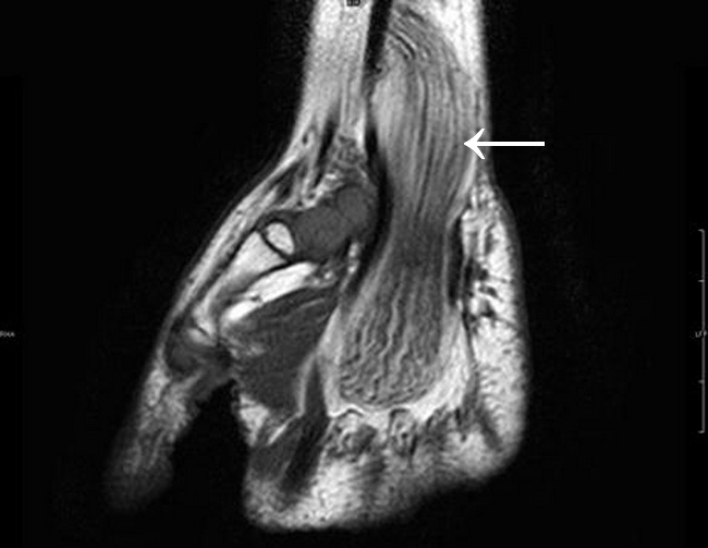
Comments:
Neural Fibrolipoma - Imaging: This MRI (Coronal T1) image is from a hand of a child. There is fusiform median nerve enlargement which is caused by thickening of nerve bundles and fibrofatty proliferation. The serpiginous low-intensity structures represent thickened nerve fascicles, surrounded by evenly distributed fat of high signal intensity on this T1-weighted image. A spaghetti-like appearance is seen in the coronal plane. The MR imaging characteristics of neural fibrolipoma are pathognomonic, obviating the need for biopsy for diagnosis.Case courtesy of Frank Gaillard, Radiopaedia.org. From the case rID: 7825
slide 4 of 9