
Extranodal Rosai-Dorfman Disease : Lung


Comments:
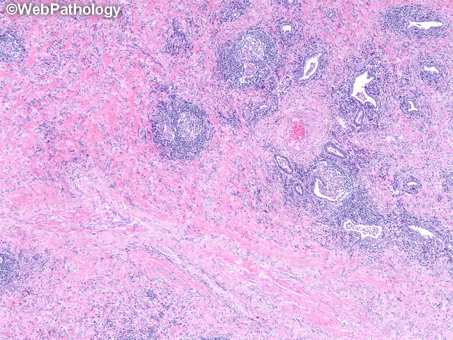
This is a case of Extranodal Rosai-Dorfman disease (RDD) involving lung. The patient was an adult female with history of persistent cough. Imaging studies showed a large right lower lobe mass involving the pleura as well as partially encasing aorta. It was considered malignant clinically. Given the diffuse nature of the mass, it was felt that it could not be resected and pneumonectomy was performed. The sections showed an inflammatory process and neoplastic entities such as sarcomatoid carcinoma or unusual sarcomas were ruled out on morphology. There is complete effacement of lung architecture by dense fibrosis, histiocytic proliferation as well as lymphoid follicles. A few residual airways can be seen on the upper right in this image. The lesional histiocytes were strongly positive for S-100 immunostain. Emperipolesis was present but not a prominent feature.