
Extranodal Rosai-Dorfman Disease : Testis
Home
 Hematopathology
Lymph Node (Non-Hematopoietic)
Non-Neoplastic Histiocytic Proliferations
Extranodal Rosai-Dorfman Disease : Testis
Hematopathology
Lymph Node (Non-Hematopoietic)
Non-Neoplastic Histiocytic Proliferations
Extranodal Rosai-Dorfman Disease : Testis

slide 45 of 53


Comments:
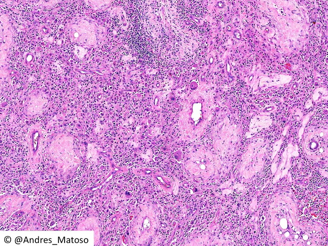
Rosai-Dorfman disease (RDD) of the testis. The image shows a mixed infiltrate of histiocytes, lymphocytes and plasma cells in the interstitium in between atrophic seminiferous tubules. Some of the histiocytes (larger multinucleated-appearing cells near the center) show emperipolesis. The differential diagnosis of testicular RDD is granulomatous orchitis, hematolymphoid disorders, germ cell tumors, and metastases. Image courtesy of: Andres Matoso, MD, Dept. of Pathology, Johns Hopkins Hospital, Baltimore, MD, USA; used with permission.
slide 45 of 53