
Metastatic Thyroid Cancers : Reporting


Comments:
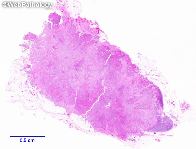
Reporting of Nodal Metastases in Thyroid Carcinomas: The pathology report should include the following elements (from AJCC Cancer Staging Manual, Eighth Edition, 2017): 1) Location of involved lymph nodes - N1a (central nodes, Level VI or VII, including pretracheal, paratracheal, prelaryngeal, or upper mediastinal) or N1b (lateral cervical lymph nodes levels I, II, III, IV, or V or retropharyngeal lymph nodes). The location information may be obtained from the operative notes. The involvement may be unilateral or bilateral. 2) Number of involved nodes with confirmed metastases. 3) Number of lymph nodes sampled. For thyroid carcinomas, there is no requirement for a minimum number of lymph nodes to be sampled. 4) Size of largest involved lymph node. 5) Size of metastatic foci within involved lymph nodes - the maximum diameter of metastatic foci in millimeters. 6) Extranodal extension of metastases by gross or microscopic examination. This low power scan of a cervical lymph node shows metastatic papillary thyroid carcinoma with extranodal extension. The tumor almost completely replaces the nodal parenchyma and spills over into surrounding adipose tissue outside the lymph node capsule. Residual lymph node can be seen on the lower right as a slightly basophilic area. Extranodal extension of metastases significantly increases the risk of persistent/recurrent disease.