
Silicone Lymphadenopathy : Differential


Comments:
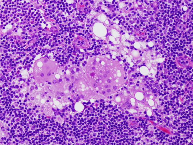
The differential diagnosis of silicone lymphadenopathy includes various causes of granulomatous lymphadenitis, lipogranuloma (coarse fat vacuoles in contrast to fine silicone vacuoles), fat necrosis (numerous foreign body giant cells), metastatic carcinomas (lobular, signet ring cell, renal cell), and signet ring cell lymphomas. Clinical history, including history of cosmetic, reconstructive or orthopedic procedure with an implant, are helpful in arriving at the correct diagnosis. The image shows an Asteroid body (center of the image) which may be seen in the cytoplasm of epithelioid or giant cells in silicone lymphadenopathy. They appear as eosinophilic, stellate or spider-like inclusions with radiating filamentous arms that contain complex lipoproteins, calcium, phosphorus, silicon, and aluminum. Besides silicone lymphadenopathy, asteroid bodies are also seen in sarcoidosis and other lymphadenopathies. Image courtesy of: Kuixing Zhang, MD; used with permission.