
PanNET : Cytology

slide 34 of 78


Comments:
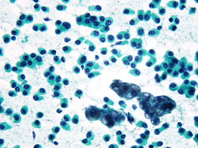
Liver Metastases from Pancreatic Neuroendocrine Tumor: This is a CT-guided FNA of a liver nodule in a 52 y/o with a mass in the tail of pancreas (same case as the previous image). There is nice contrast between discohesive tumor cells and clusters of benign hepatocytes. Final diagnosis: Metastatic Pancreatic Neuroendocrine Tumor. Image courtesy of: Syed Z. Ali, MD; Director of Cytopathology, Johns Hopkins University, Baltimore, Maryland, USA; used with permission.
slide 34 of 78