
Melanoma Immunohistochemistry : Melan-A


Comments:
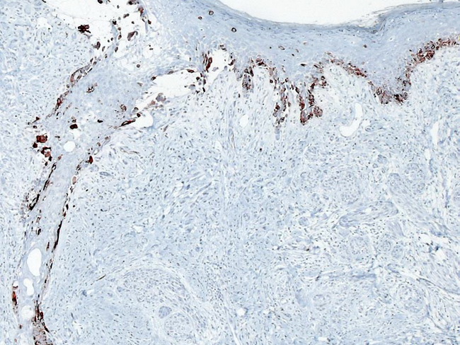
Immunohistochemical stains play a vital role in the diagnosis of amelanotic, spindle cell, and epithelioid variants of melanoma and their distinction from poorly-differentiated carcinomas as well as mesenchymal tumors. A classic case of melanoma is immunoreactive for S-100 protein, HMB-45, Melan-A, tyrosinase, Microphthalmia Transcription Factor (MITF), and vimentin. Melan-A (MART-1) is a melanosomal differentiation antigen that is expressed by melanomas, PEComas, adrenocortical, Leydig cell, Sertoli cell, and granulosa cell tumors. It is a highly sensitive marker for epithelioid melanomas (positive in 80% of cases) but is less commonly positive in spindle cell and desmoplastic melanomas. Reduced expression of MART-1 is an adverse prognostic indicator and correlates with thick melanomas, reduced disease-free survival, and increased patient mortality. The image shows Melan-A positivity in the intraepidermal component of a lentigo maligna melanoma. The invasive dermal component was desmoplastic melanoma and was negative for Melan-A but strongly positive for S-100 protein. Breslow thickness was > 9mm. Clark level 5. Image copyright: pathorama.ch.