
Urothelial Carcinoma-in-situ

slide 7 of 22


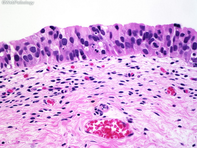
Comments:
There is severe cytologic atypia consisting of loss of polarity, nuclear enlargement and hyperchromasia, and nuclear pleomorphism. Apoptotic cells and mitotic figures are readily seen. The urothelial thickness is decreased and the cytologic atypia involves all layers. The lamina propria shows numerous capillary-caliber vessels and this feature is responsible for the erythematous appearance of CIS on cystoscopy.
slide 7 of 22