
Ganglioneuroma

slide 7 of 27


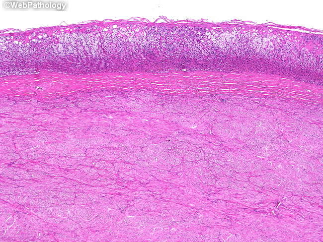
Comments:
Ganglioneuromas usually present as large masses in the posterior mediastinum (40% of cases) or retroperitoneum (30% of cases). Although adrenal involvement (shown here) is comparatively less common (20% of cases), it must be considered during the evaluation of an adrenal mass. The image shows a thin rim of stretched out adrenal cortex overlying the tumor. The tumor is well-circumscribed and contains a fibrous capsule.
slide 7 of 27