
FAP : Microscopic Features


Comments:
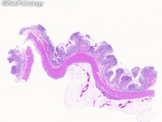
Microscopic Features of Familial Adenomatous Polyposis (FAP): All types of adenomatous polyps are seen, including tubular, tubulovillous, and villous adenomas. The gross pathology and microscopic features are identical to the sporadic adenomas as described previously. This whole slide scan shows numerous small polyps which appear basophilic due to low-grade dysplasia in comparison to the the surrounding normal mucosa. The lesions begin with monoclonal expansion of a single abnormal cell which gives rise to a unicryptal adenoma. The proliferative compartment spreads throughout the crypt instead of being confined to the crypt base. The proliferating cells at the surface are not shed into the lumen due to failure of apoptosis. With the accumulation of proliferating cells, additional adenomatous glands are created either by infolding or by branching, thereby producing macroscopic lesions. As the adenoma enlarges, the neoplastic cells become polyclonal and acquire additional mutations.