
Mesemchymal Chondrosarcoma : Morphology


Comments:
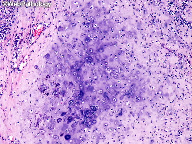
Microscopic Features: This image shows a chondroid focus undergoing calcification (dark blue/purple areas) in a mesenchymal chondrosarcoma (MC). Chondroid foci in MC are often calcified or ossified. Osteoid trabeculae or delicate, lace-like osteoid may be seen between the small tumor cells. The relative proportion of the two components varies from case to case. Some cases have large islands of well-differentiated hyaline cartilage and a minor component of small, undifferentiated blue cells squeezed in between. Such cases closely resemble conventional chondrosarcoma. Other cases are composed predominantly of sheets of small round malignant cells and have small foci of barely perceptible chondroid matrix. Such cases can mimic malignant lymphoma or Ewing sarcoma.