
Solid Pseudopapillary Tumor : Radiology


Comments:
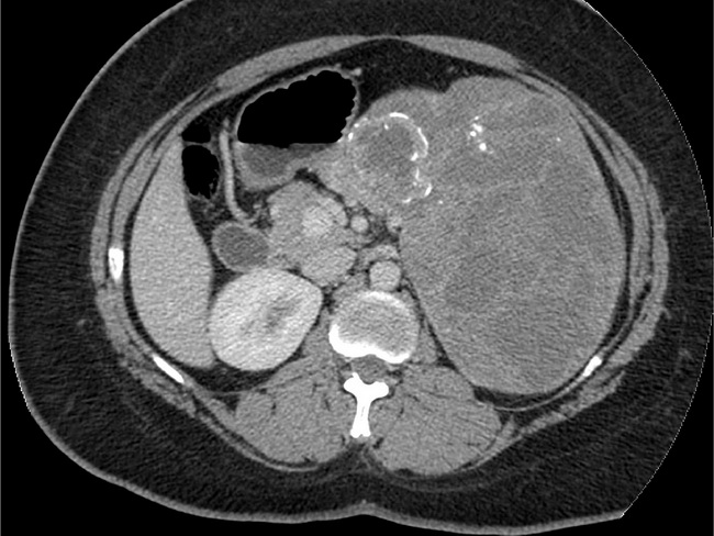
On ultrasound and computed tomography (CT), solid pseudopapillary tumor (SPT) of pancreas appears as a large, sharply demarcated, heterogenous mass with solid and cystic components. Areas of hemorrhage and calcification are frequently seen. With contrast, there is peripheral enchancing of the solid areas. Endoscopic ultrasonography-guided FNA has been increasingly used to diagnose SPT preoperatively. FNA smears are cellular and show delicate, branching papillary fronds lined by bland, monomorphic cells with round to oval nuclei.This CT abdomen (with contrast) of SPT is from a 25 y/o female who presented with bloating, early satiety, and weight loss. It shows a large well-defined encapsulated heterogeneous mass in pancreatic body/tail. Additional findings included: calcifications, cystic/necrotic foci, peripheral and septal enhancement, and vascular and visceral displacement. There were no metastases. Case courtesy of Dr Jeremy Frank, Radiopaedia.org. From the case rID: 32680