
Sertoli Cell Tumor

slide 48 of 104


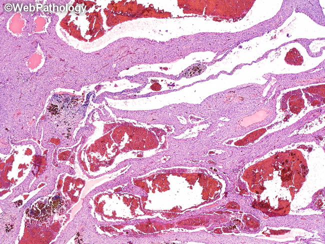
Comments:
This Sertoli cell tumor had a somewhat atypical presentation. It was a well-circumscribed multicystic hemorrhagic mass. There was no appreciable tubular differentiation. The diagnosis was confirmed with immunohistochemical features: positive for calretinin, CD99, nuclear beta-catenin, WT!, and vimentin. The tumor was negative for inhibin-alpha - a feature seen in about 50% of Sertoli cell tumors.
slide 48 of 104