
Epidermodysplasia Verruciformis

slide 45 of 56


Comments:
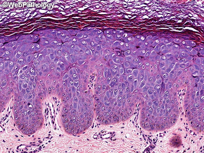
The image shows typical histologic features of epidermodysplasia verruciformis. There is acanthosis, hyperkeratosis, and hypergranulosis. Keratinocytes in the upper layers of epidermis show enlarged nuclei and voluminous cytoplasm with bluish-gray pallor.
slide 45 of 56