
Metaplastic Meningioma

slide 43 of 60


Comments:
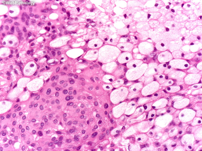
On the left is a syncytium of bland looking meningothelial cells with ample pink cytoplasm and uniform nuclei. The right side of the image shows cytoplasmic xanthomatous change due to fat accumulation and vacuolization (right upper corner).
slide 43 of 60