
Post-radiation Angiosarcoma

slide 43 of 115


Comments:
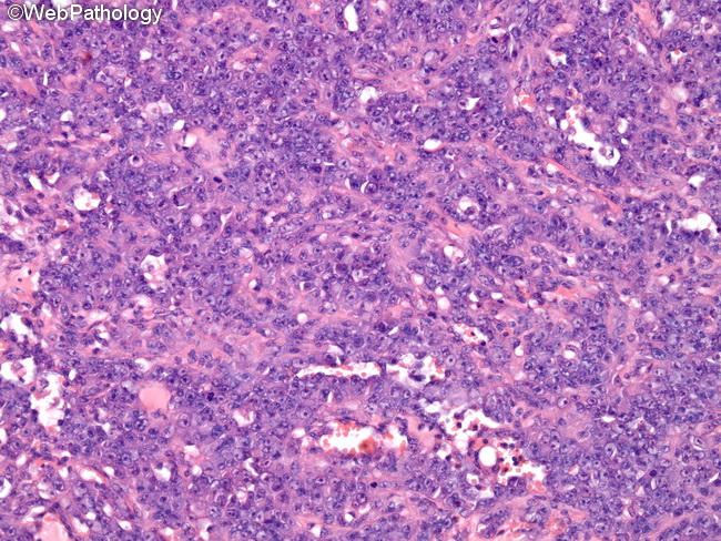
The image shows epithelioid areas in a post-radiation angiosarcoma of the breast (same case as the previous two images). The tumor cells in this focus are arranged in solid sheets punctuated by a few vascular channels. There is moderate amount of eosinophilic cytoplasm, large vesicular nuclei, prominent nucleoli, and increased mitotic activity. Such areas, on small core biopsy specimens, can be misdiagnosed as carcinomas.
slide 43 of 115